CARDIOVASCULAR ORGANS (ANATOMICAL MICROSCOPY)
14.1
Aorta
Specimen:
Specimen Details:
Organ: Aortic wall
Origin: Monkey
Staining: RFAL
Method and Specimen Description:
Normal histological sections were prepared and stained with RFAL, a method that vividly demonstrates elastic fibers within the vessel wall. The tissue originates from a region close to the heart — an area involved in the Windkessel effect (pressure reservoir function of the aorta).
Objective of the Examination:
To study the elastic wall structure of an artery, to identify its individual layers, and to recognize other incidentally sectioned structures.
Special Features of the Specimen:
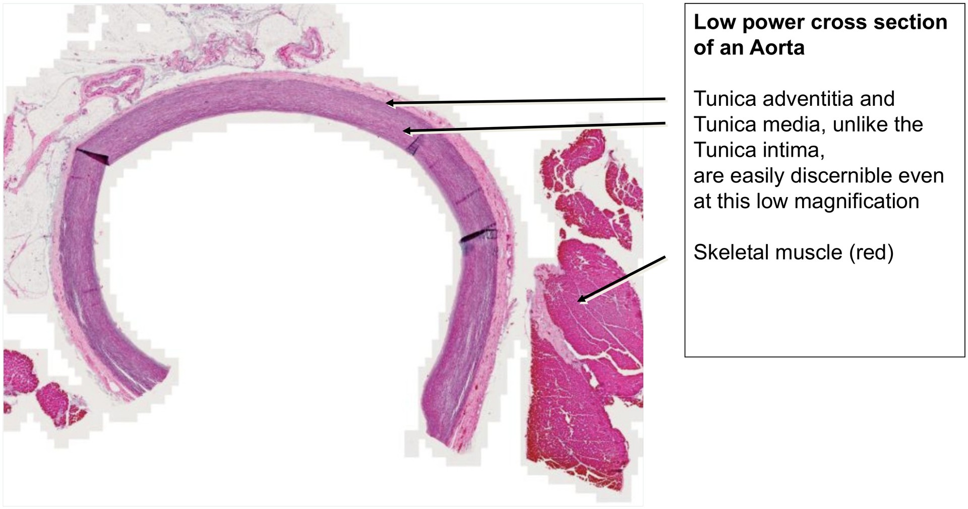
The aorta, like all arteries, possesses a three-layered wall structure clearly visible at low magnification:
- Tunica intima
- Tunica media
- Tunica adventitia
In this section, minor folding artefacts may be observed, resulting from the cutting process.
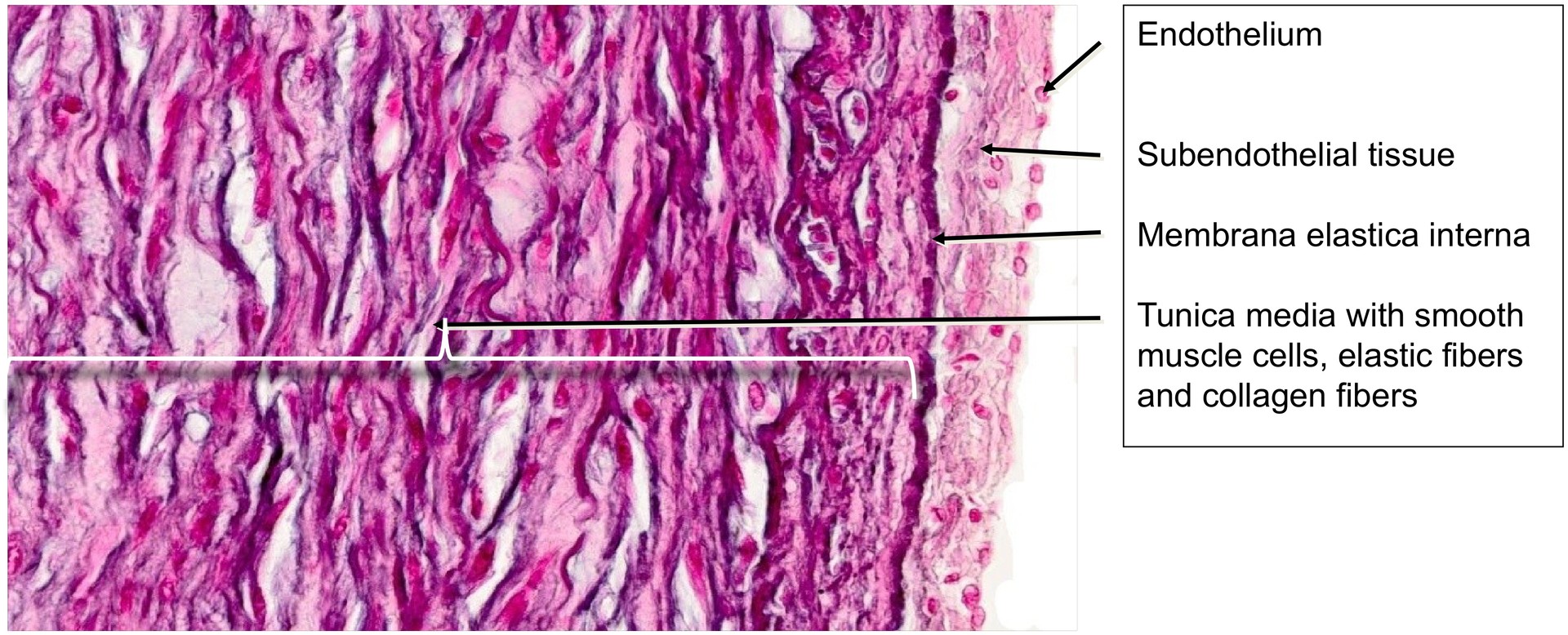
Tunica intima:
- Lined by a delicate endothelium, not preserved in all regions.
- Beneath it lies the subendothelial layer (stratum subendotheliale), which may contain occasional longitudinally oriented smooth muscle cells.
- The internal elastic lamina demarcates the border between the intima and media and is distinctly visible as a strong elastic layer.
Tunica media:
- Composed of circularly arranged smooth muscle cells interspersed with elastic and collagen fibers arranged in concentric sheets.
- RFAL staining highlights the elastic fibers particularly well.
- An external elastic lamina is often indistinct or absent in the aorta.
- The transition to the tunica adventitia is clearly defined by the disappearance of smooth muscle cells and the appearance of larger elastic fibers.
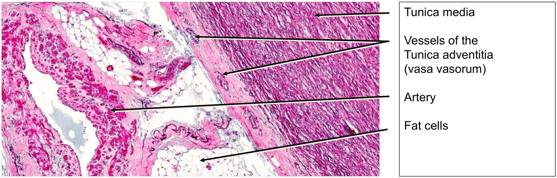
Tunica adventitia:
- Contains numerous vasa vasorum, collagen fibers, and fat cells.
- Represents the outermost supportive layer of the vessel.
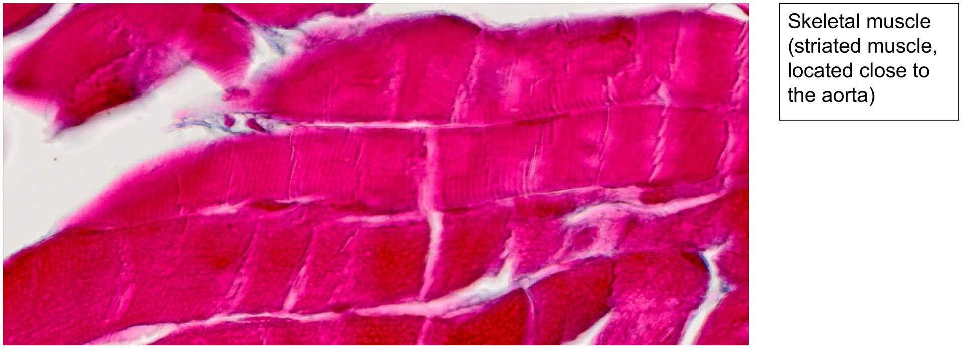
- May also include co-sectioned connective tissue, adipose tissue, and skeletal muscle fibres at the periphery of the specimen.
Tasks:
- Identify and delineate the individual layers of the aortic wall: tunica intima, tunica media, and tunica adventitia.
- Locate regions with intact endothelium and examine the structure of the subendothelial layer.
- Trace the internal elastic lamina over a longer distance and compare it with the elastic fibers within the tunica media.
- Assess the ratio of elastic fibers to smooth muscle cells in the aortic wall.
- Locate several vasa vasorum and fat cells within the tunica adventitia.
- Examine the peripheral regions of the specimen and identify any co-sectioned structures (e.g. fat tissue, vessels, skeletal muscle).

License
University of Basel
Downloads
- Hide »
- Toggle Theme
-
History
/en/histology-12/cardiovascular-organs-anatomical-microscopy-50/aorta-312 -
Versions
Django 5.2.14 -
Time
CPU: 174.85ms (178.08ms) - Settings
- Headers
-
Request
apply_cache -
SQL
0 queries in 0.00ms -
Static files
7 files used -
Templates
step/step_home.html -
Cache
56 calls in 18.44ms -
Cachalot
Last invalidation: 5 hours, 28 minutes -
Signals
129 receivers of 15 signals -
Intercept redirects
-
Profiling