CARDIOVASCULAR ORGANS (ANATOMICAL MICROSCOPY)
14.2
Artery and vein
Specimen:
Specimen Details:
Organ: Artery and Vein
Origin: Human
Staining: RFAL
Method and Specimen Description:
Normal histological sections were stained with RFAL, which distinctly highlights the elastic fibres within the vessel walls.
Objective of the Examination:
- To understand the structural differences between arteries and veins.
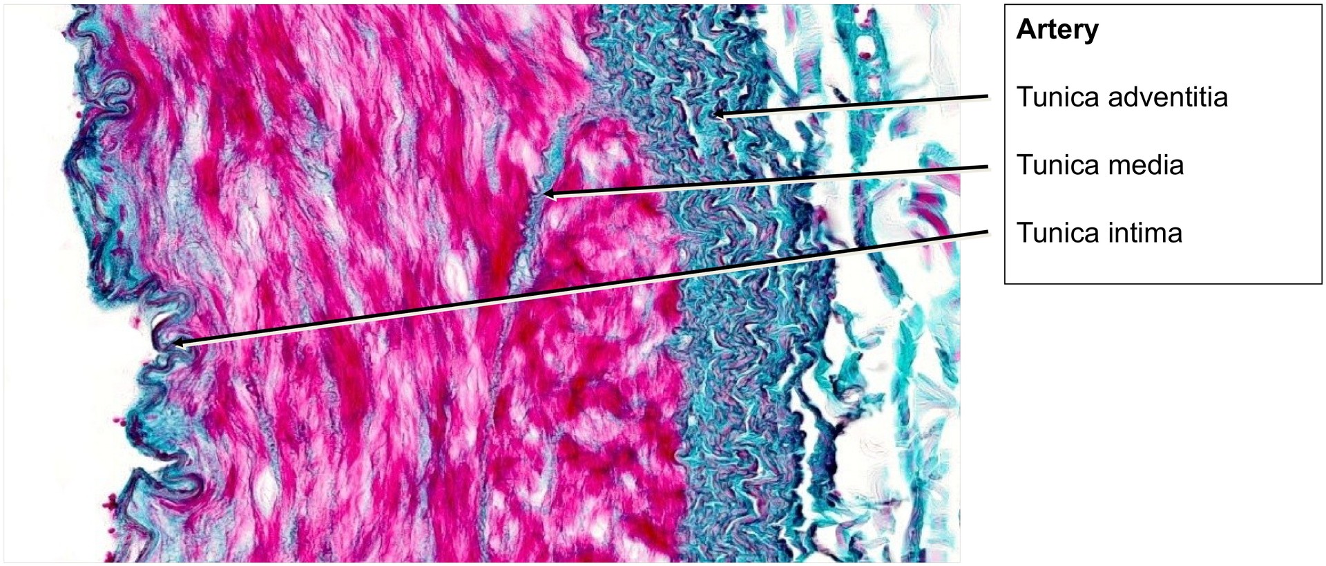
- To study the wall structure of a muscular artery.
Special Features of the Specimen:
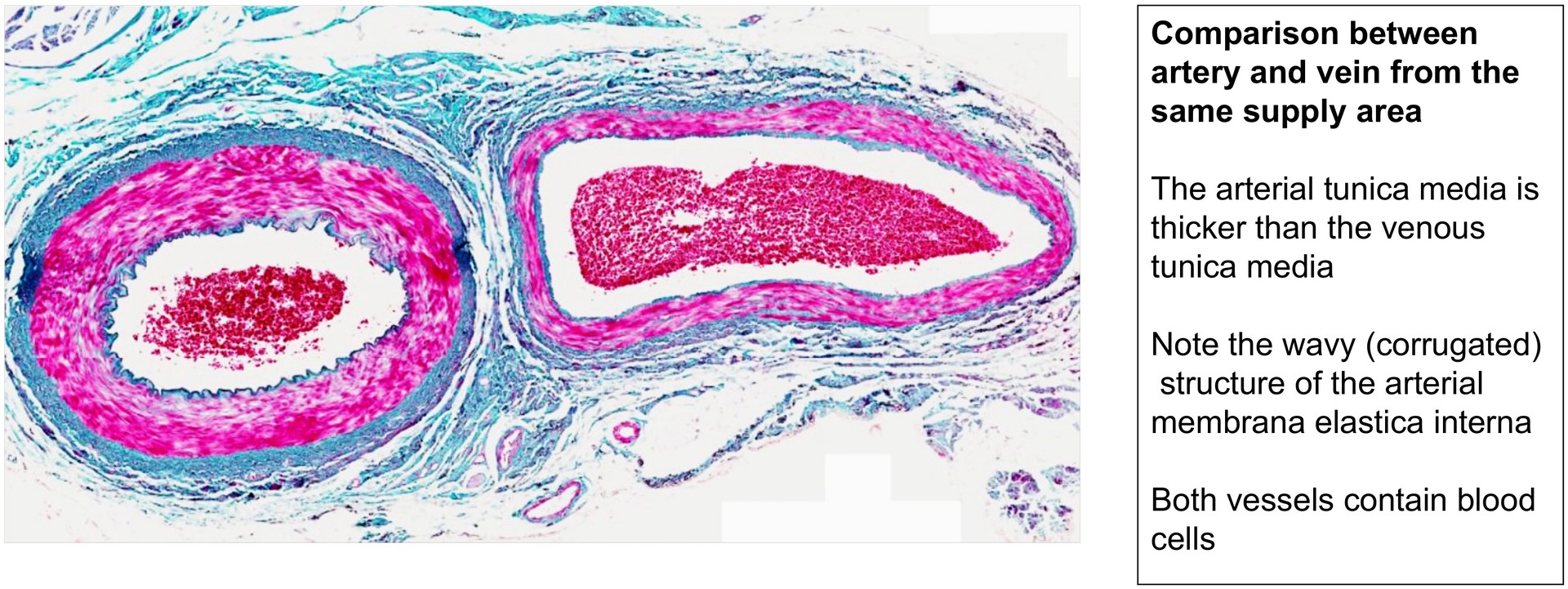
Even under low magnification, a marked difference between the artery and the vein is evident: the arterial wall is considerably thicker than the venous wall. However, this rule applies only to vessels of the same drainage area; in some regions, a vein may possess a thicker wall than an artery from another area.
Artery:
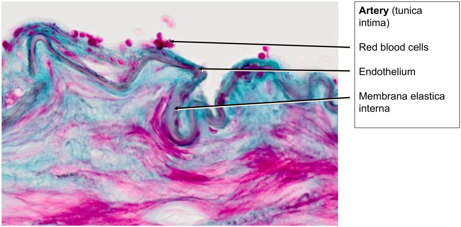
- The endothelium lies directly on the internal elastic lamina; a subendothelial layer is only weakly developed (best seen at high magnification).
- The internal elastic lamina appears strongly convoluted due to contraction of the muscular tunica media during fixation.
- The media consists of smooth muscle cells interspersed with a few elastic fibres, markedly fewer than in the aorta.
- The adventitia is thinner and less distinct than that of the aorta, containing small connective tissue elements and vasa vasorum in some areas.
Vein:
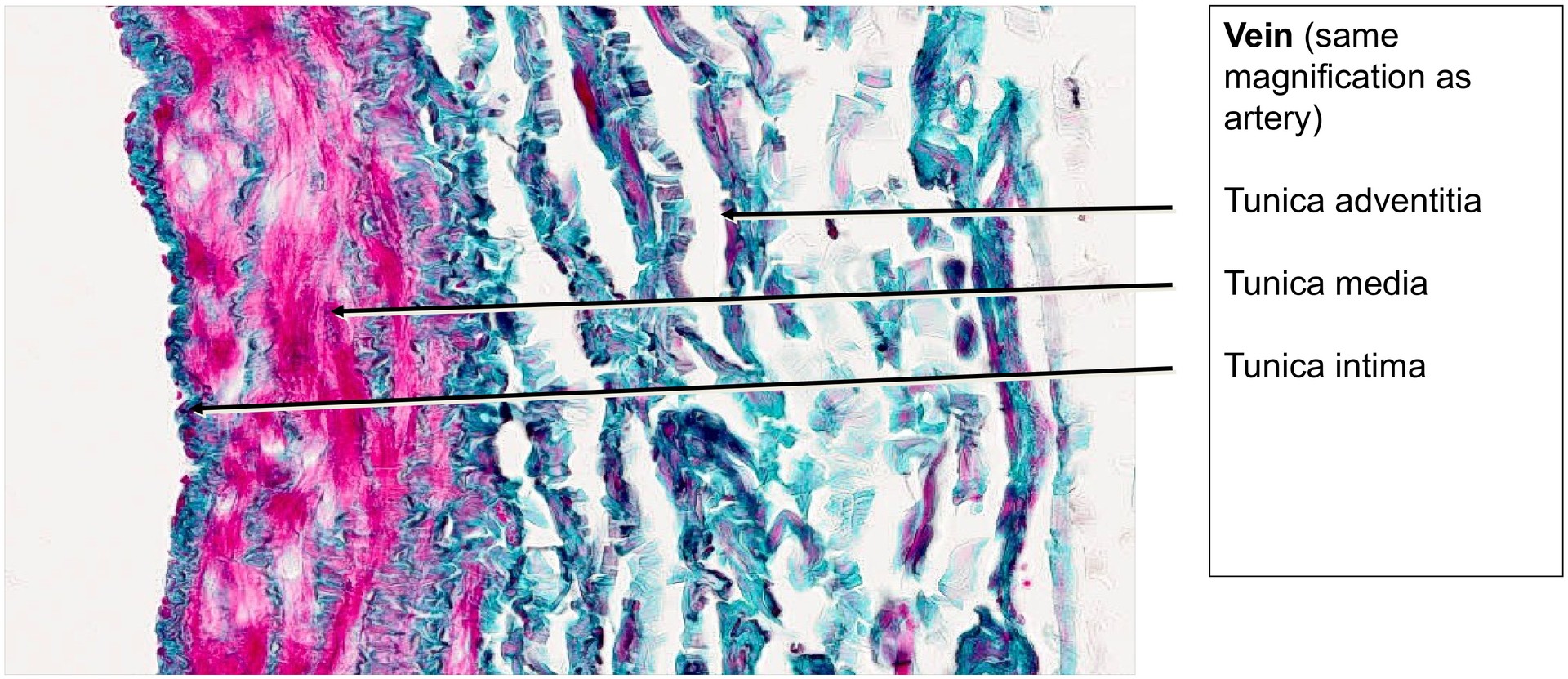
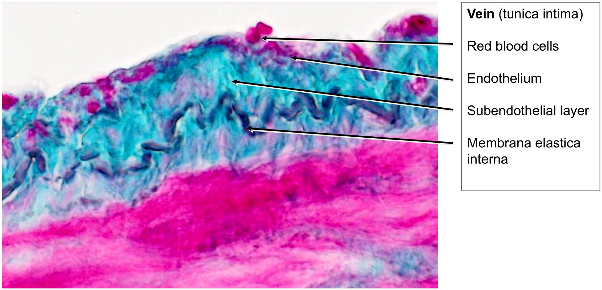
- The endothelium is clearly visible, and a subendothelial layer is present.
- The tunica media is relatively narrow, containing fewer smooth muscle cells and only sparse elastic fibers.
- The internal elastic lamina is thin and less corrugated, reflecting the weaker muscular component.
- The tunica adventitia is poorly defined, merging gradually into the surrounding connective tissue.
Tasks:
- At low magnification, identify and distinguish the artery and vein.
- Compare the muscular thickness of the tunica media in both vessels.
- Evaluate the presence and arrangement of elastic fibers and the internal elastic lamina in each vessel.
- Examine the smaller vessels in the vicinity of the larger ones.
- Locate and identify the nerve cross-section visible within the specimen.
License
University of Basel
Downloads
- Hide »
- Toggle Theme
-
History
/en/histology-12/cardiovascular-organs-anatomical-microscopy-50/artery-and-vein-313 -
Versions
Django 5.2.14 -
Time
CPU: 163.72ms (166.53ms) - Settings
- Headers
-
Request
apply_cache -
SQL
0 queries in 0.00ms -
Static files
7 files used -
Templates
step/step_home.html -
Cache
53 calls in 16.94ms -
Cachalot
Last invalidation: 1 hour, 8 minutes -
Signals
129 receivers of 15 signals -
Intercept redirects
-
Profiling
DJDT