NERVOUS SYSTEM (ANATOMICAL MICROSCOPY)
16.5
Cerebellum 1
Specimen:
SPECIMEN DETAILS:
Organ: Cerebellum
Source: Human
Staining: Hematoxylin and Eosin (H&E)
METHOD AND SPECIMEN DESCRIPTION:
This is a normal section of the adult human cerebellum, stained with Hematoxylin and Eosin (H&E).
- Hematoxylin stains cell nuclei blue, highlighting chromatin and nucleoli.
- Eosin stains cytoplasm and extracellular matrix pink to red.
Only a small portion of the cerebellum is visible in the section, as the organ’s overall size and folding pattern prevent inclusion of the entire structure on a single slide.
OBJECTIVE OF THE EXAMINATION:
To study the layered structure of the cerebellar cortex and recognise the main neuronal and glial cell types.
Special Features of the Specimen:
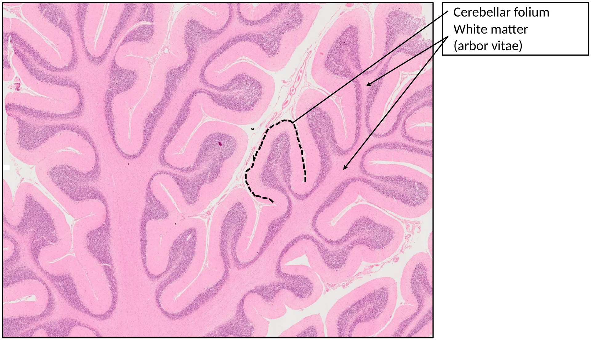
The cerebellum forms numerous densely packed folds (folia), which together produce the characteristic arbor vitae or “tree of life” appearance in cross-section.
At medium magnification, a clear distinction between the cortex and the underlying white matter (medulla) can be observed.
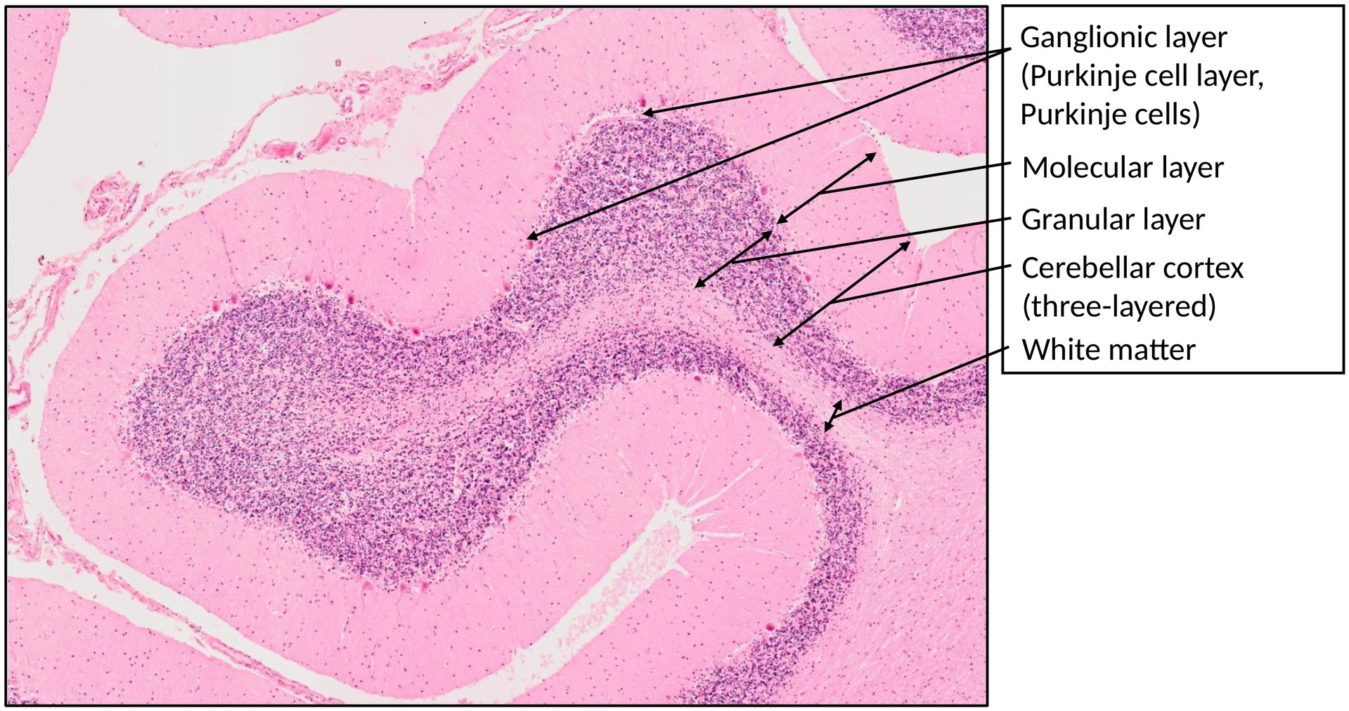
Cerebellar Cortex:
The cerebellar cortex is three-layered, arranged as follows (from superficial to deep):
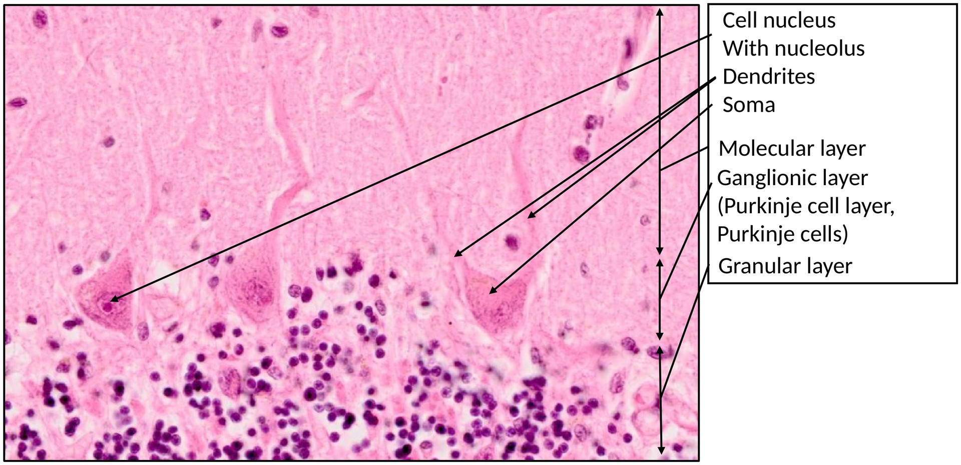
- Molecular layer –
- Contains loosely arranged neurons, primarily stellate cells (superficially) and basket cells (near the Purkinje cell layer).
- Rich in unmyelinated axons and dendritic arbors of Purkinje cells.
- Ganglionic (Purkinje cell) layer –
- A single row of large Purkinje cell bodies, each with a distinctive flask-shaped soma.
- Primary dendrites extend into the molecular layer and form elaborate arborizations.
- Granular layer –
- Densely packed with small granule cells, whose axons ascend to the molecular layer, forming parallel fibers.
- Larger Golgi cells are scattered near the Purkinje cell layer.
- Bergmann glial cells, whose somata lie near the Purkinje cells, are present but not clearly distinguishable in H&E preparations.
White Matter:
- Appears paler due to dissolution of myelin during processing.
- Contains axons of Purkinje cells, as well as the cell bodies of oligodendrocytes, astrocytes, and endothelial cells.
TASKS:
- To which structures do the Purkinje cells project?
- From where do the moss fiber afferents reach the granule cells?
- From where do the climbing fiber afferents reach the Purkinje cells?
- Approximately how many granule cells are present in the human cerebellum?
License
University of Basel
- Hide »
- Toggle Theme
-
History
/en/histology-12/nervensystem-anatomische-mikroskopie-52/cerebellum-1-335 -
Versions
Django 5.2.14 -
Time
CPU: 164.27ms (167.31ms) - Settings
- Headers
-
Request
apply_cache -
SQL
0 queries in 0.00ms -
Static files
7 files used -
Templates
step/step_home.html -
Cache
53 calls in 17.55ms -
Cachalot
Last invalidation: 2 hours, 7 minutes -
Signals
129 receivers of 15 signals -
Intercept redirects
-
Profiling